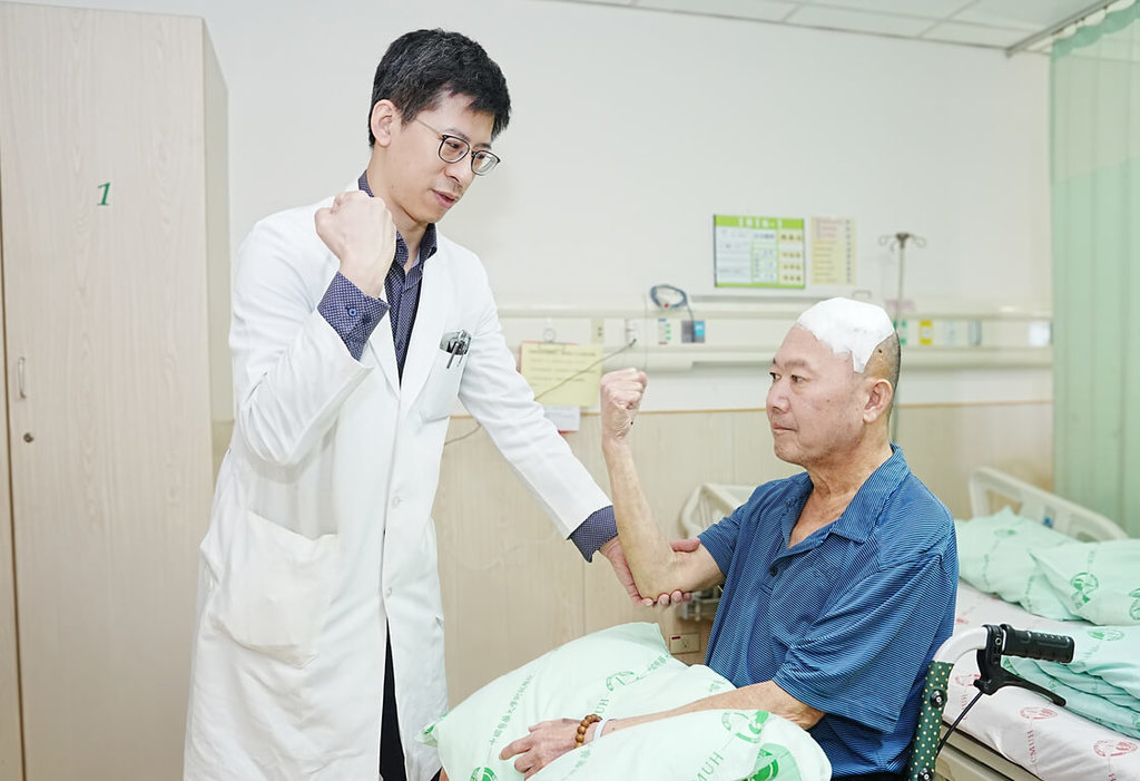
TAICHUNG, Taiwan (CNA) -- A patient in Taichung with a history of stroke suddenly found himself unable to get up on his own. Medical examinations revealed a brain tumor pressing against the motor cortex, leading the medical team to perform awake craniotomy surgery. By identifying the safe range for tumor removal, the patient was able to regain the ability to walk and perform daily activities after the operation.
China Medical University Hospital announced in a press release today that Mr. Li, a 60-year-old patient with a mild history of stroke, had gradually experienced weakness in his right limbs over the past two months. One night, he fell and was unable to get up on his own. He was rushed to the emergency room, where examinations diagnosed him with a falx meningioma adjacent to the left motor area. The team performed awake craniotomy surgery to remove the tumor. Post-surgery, Mr. Li's right limb weakness significantly improved, and he regained the ability to walk and perform daily activities, leading to a smooth discharge and return home.
Dr. Hsu Wei-lin, a neurosurgeon at CMUH, stated that Mr. Li's tumor was located close to the cerebral motor cortex and important descending motor nerve fibers. If traditional general anesthesia had been used for the surgery, it would have been difficult to determine the functional boundaries in real-time during the operation, potentially increasing the risk of postoperative limb weakness or even paralysis.
Dr. Hsu mentioned that the team adopted an awake craniotomy strategy. Under anesthesia and neurological monitoring, the patient remained awake during critical surgical stages. Through intraoperative cortical and subcortical electrical stimulation for localization, and simultaneous monitoring of hand and lower limb motor responses, as well as continuous testing of language and motor functions, the safe range for tumor removal was identified with the aid of real-time functional localization. This allowed for the maximum possible tumor resection while avoiding damage to critical motor neural pathways.
Additionally, Dr. Hsu's medical team had previously performed awake craniotomy surgery on a 56-year-old patient with recurrent glioblastoma multiforme. After the tumor recurred, it had spread along the subcortical white matter nerve fibers of the brain and began to affect language function.
Dr. Hsu pointed out that high-grade gliomas like this one do not have clear boundaries, and surgery requires a balance between reducing tumor burden and preserving language, cognitive, and spatial functions. In addition to traditional motor and language tests, the team also incorporated advanced functional tests such as cognition, calculation, and spatial cognition. Through real-time functional monitoring and subcortical fiber stimulation, they aimed to protect important neural circuits as much as possible. The patient did not experience any new neurological deficits after the surgery and was smoothly discharged one week later. (Editor: Chen Ching-fang) 06/24/115
Choose to stand with the facts. Your every donation is a force to protect the freedom of the press.
Download the CNA "One-Stop News" APP to get the latest news in real-time.
The text, images, and videos on this website may not be reproduced, publicly broadcast, publicly transmitted, or used without authorization.
FACT BOX
- Source: CNA (Central News Agency)
- Category: 医療